
More Info
Script
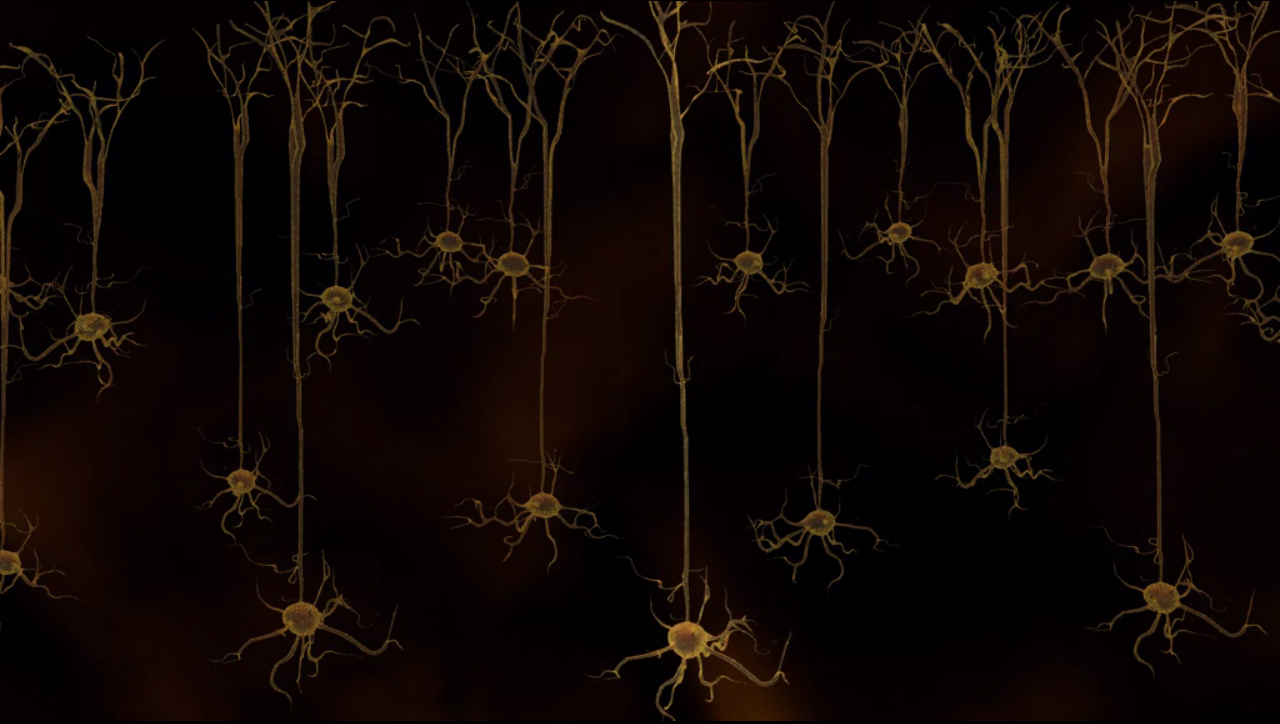
Structurally exquisite computational wonders, the billions of neurons in your brain control the body and constitute the mind. This areal view shows the outer most shell of the brain: the cerebral cortex. Neurons of many different shapes and sizes that perform different functions populate specific areas of the cortex. The slender and long protrusions that extend towards the surface are the dendritic trees of neurons. Their task is to collect small packets of information from other neurons and funnel it to the cell body. These packets can excite or inhibit, but when excitation exceeds inhibition an action potential is produced by the neuron's cell body. Action potentials are one of the most important means of communication between neurons. This electrical signaling is possible because neurons have excitable membranes thanks to the voltage difference across their membrane and ion selective pores. These properties allow neurons to carry signals over long distances via their axons. These fast conducting wires carry action potentials away from the cell body. Not all neurons communicate with each other; instead, specific patterns of connections arise through development and experience. When neurons do communicate, they form a structure called a synapse: where the axon terminal of one neuron meets the dendritic tree of a different neuron. Let’s take a closer look at what happens in this critical communication event called synaptic transmission. Action potentials are propagated by axons and upon reaching the axon terminal voltage sensitive channels on the membrane open and allow the entry of calcium ions exclusively. Calcium is an important intracellular messenger: once it enters the axon terminal calcium ions trigger a cascade of events that lead to the release of neurotransmitters: the central event of synaptic transmission. Neurotransmitters are small molecules cells use to communicate with each other. In the axon terminal they are packaged in vesicles where they are protected from damage and their concentration and release can be tightly regulated. Vesicles have a calcium-sensing protein called synaptotagmin. Once calcium binds to synaptotagmin SNARE is activated; this protein complex is responsible for fusing the synaptic vesicle and cell membranes. Once fusion occurs, a pore opens and the neurotransmitter diffuses into the synaptic cleft. This process is very fast: neurotransmitter release occurs less than 0.5 millisecond after calcium enters the terminal. Neurotransmitter release is only half of the story of synaptic transmission. On the opposite side is the postsynaptic membrane: the receiver of the message, and its task is to convert a chemical message into an electrical signal. The postsynaptic membrane has channels that recognize specifically the neurotransmitter released and allow the passage of specific ions only when the neurotransmitter is attached. The charge of the ion allowed to enter the postsynaptic membrane determines whether the signal will excite or inhibit the neuron. These electrical signals are smaller than action potentials and constitute the small packets of information collected by dendritic trees where the process of integrating signals and synaptic transmission begins once again.
To learn more about this topic please contact Professor Hysell Oviedo: hoviedo@ccny.cuny.edu CCNY
Subject
Biology
Type
Animation
Format
MPEG-4
Date
2015
Contributor
Advait Apte, Ching-Jung Chen, Katie Cheng, Dalia Gracia, Lenn Hypolite, Rafay Malik, Helena Marvin, Hysell Oviedo.
Publisher
The City College Libraries, New York, New York
Identifier
ANI008
Language
English
Rights
Synaptic Transmission by City College of New York Digital Scholarship Services is licensed under a Creative Commons Attribution 4.0 International License.